Hypokalemia is a medical condition that affects dogs and cats, and is characterized by abnormally low serum potassium ion (K+) concentration. The normal reference interval for serum K+ concentration in dogs and cats is 3.5 to 5.5 mEq/L (mmol/L). If the serum K+ concentration falls below 3.5 mEq/L (mmol/L), the patient is said to be hypokalemic.
Estimated Serum K+ Concentration Reference Intervals in Dogs and Cats:
Normal: 3.5 to 5.5 mEq/L (mmol/L)
Hypokalemia: <3.5 mEq/L (mmol/L)
Mild hypokalemia: 3 to 3.4 mEq/L (mmol/L)
Moderate hypokalemia: 2.5 to 2.9 mEq/L (mmol/L)
Severe hypokalemia: <2.5 mEq/L (mmol/L)
Note that the above are estimates and that reference intervals vary slightly between labs and sources.
Introduction
K+ is an electrolyte, more specifically a cation (a positively charged ion), that plays an essential role in many body functions. It is the body’s major intracellular cation, with ~95 to 98% of total body K+ being located within the intracellular fluid (ICF) compartment. K+ is responsible for intracellular fluid (ICF) volume maintenance, as well as essential for many body functions. The intracellular fluid (ICF) compartment to extracellular fluid (ECF) compartment ratio of K+ concentration is a major determinant of resting cell membrane potential. This K+ concentration gradient is principally maintained by Na+/K+-ATPase in cell membranes, which pumps out 3 Na+ for every 2 K+ into the cell. Total body K+ is primarily regulated by the kidneys, mainly via aldosterone , which stimulates Na+ reabsorption and K+ excretion. The GI tract primarily non-selectively absorbs and excretes K+. Note that serum K+ concentration does not always reflect total body K+.
Hypokalemia causes hyperpolarization of the resting cell membrane potential. Systems affected by hypokalemia include neuromuscular (nerve, skeletal muscle (including muscles of respiration), cardiac muscle, smooth muscle), renal, and metabolic.
Causes
There are several causes of hypokalemia in dogs and cats, including:
- Increased K+ loss: increased renal (e.g. chronic kidney disease, hypokalemic nephropathy, renal tubular acidosis, renal failure, postobstructive diuresis, IV fluid diuresis, diabetic ketoacidosis, hyperaldosteronism, hyperadrenocorticism, hypomagnesemia, loop diuretic, thiazide diuretic, amphotericin B, penicillins, glucocorticoid excess) or gastrointestinal (e.g. vomiting, diarrhea, obstruction) K+loss.
- Translocation of K+ between fluid compartments (from the extracellular fluid (ECF) compartment into the intracellular fluid (ICF) compartment): insulin, catecholamine release (e.g. epinephrine), glucose, sodium bicarbonate, alkalemia, beta2-adrenergic agonist (e.g. albuterol, terbutaline), rattlesnake envenomation, hypothermia, endotoxemia, familial disorder in Burmese cats (hypokalemic periodic paralysis).
- Iatrogenic: K+-deficient diet, K+-deficient IV fluid administration, loop diuretic, thiazide diuretic, amphotericin B, penicillins, insulin, epinephrine, glucose, sodium bicarbonate, beta2-adrenergic agonist (e.g. albuterol, terbutaline).
- Decreased K+ intake: dietary (anorexia or starvation, K+-deficient diet), K+-deficient IV fluid administration, bentonite clay ingestion (e.g. clumping cat litter).
- Artifact: hyperlipidemia.
Clinical Signs
Clinical signs of hypokalemia vary depending on the severity of the condition. In mild cases, hypokalemia may be asymptomatic. In moderate to severe cases, the following symptoms may be present:
- Mild hypokalemia: Typically presents asymptomatic.
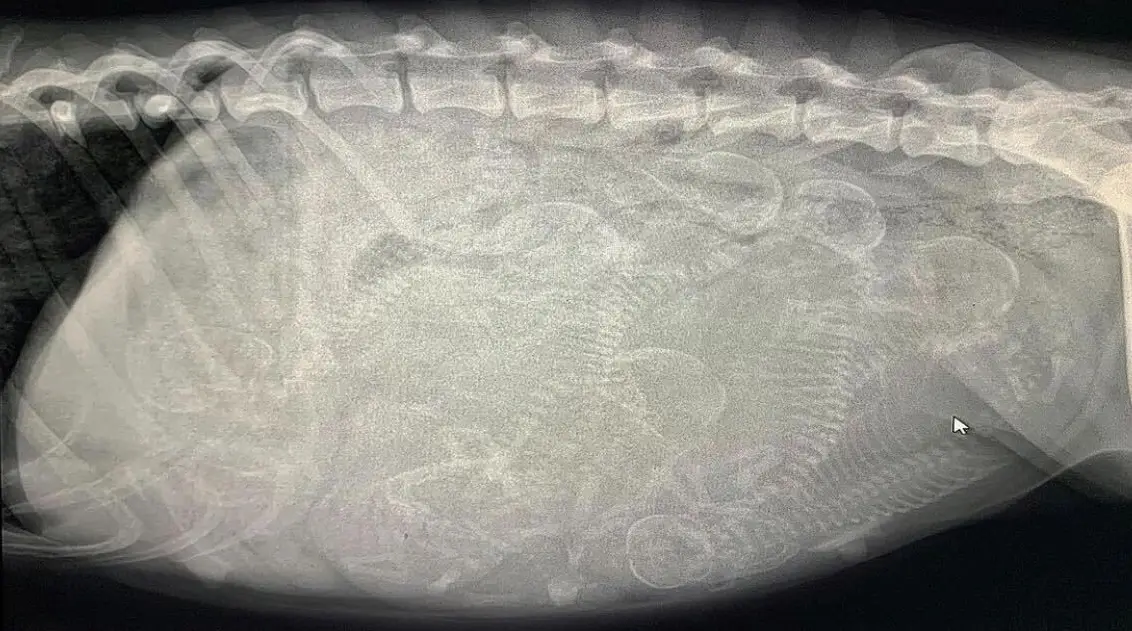
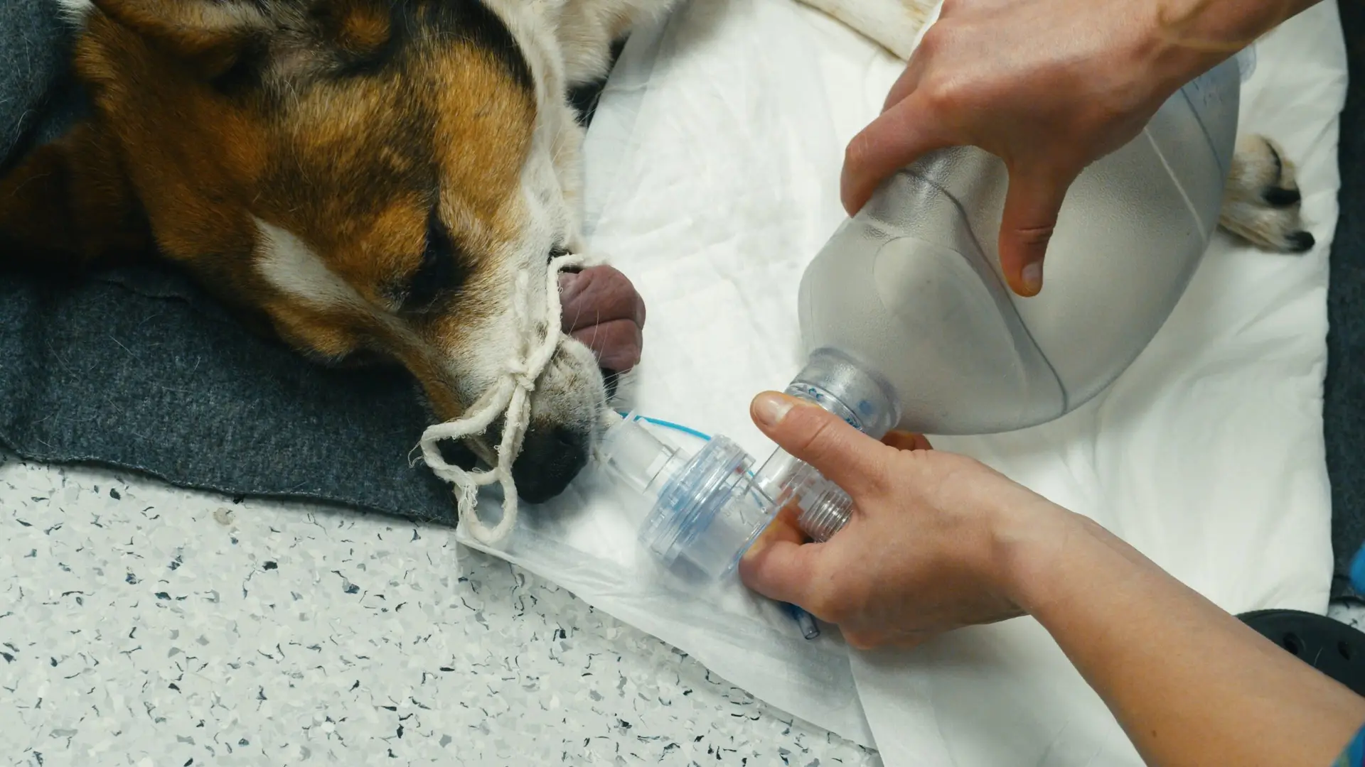
- Moderate to severe hypokalemia: Clinical signs include anorexia, vomiting, diarrhea, weight loss, lethargy, confusion, skeletal muscle weakness (ventroflexion of the neck, forelimb hypermetria, broad-based hind limb stance, hind limb weakness, paresis, paralysis, respiratory muscle paralysis, rhabdomyolysis), cardiac arrhythmias, renal (polyuria, polydipsia), smooth muscle dysfunction (constipation due to decreased intestinal motility), metabolic (acid-base balance, glucose homeostasis).
Typical Treatment Options
Identify and treat the underlying disease process, as well as provide K+ supplementation.
Mild hypokalemia: Oral K+ supplementation with potassium gluconate (e.g. Tumil-K) or potassium citrate -> dosage 5 to 1 mEq/kg PO every 12 to 24 hours, can be mixed into food. Prescription renal diets are typically formulated with supplemented K+. Good sources of K+ include bananas and kiwis.
Moderate to severe hypokalemia: With moderate (not severe) hypokalemia, if the patient is eating and drinking, not vomiting, and relatively stable clinically, then inpatient PO supplementation with frequent monitoring may also be an option. Parenteral (usually IV) K+ supplementation. The supplement of choice is potassium chloride (KCl) added to the fluid bag -> the usual CRI dosage for K+ supplementation is 0.1 to 0.2 mEq/kg/hr IV. The dosage should not exceed 0.5 mEq/kg/hr IV (which is known as Kmax), except in some very dire circumstances where increasing the dosage up to 1 to 1.5 mEq/kg/hr IV may be considered with close ECG monitoring. Always add KCl to the fluid bag, and never administer KCl as a sole agent. Mix the fluid bag extremely well. Refer to our “Estimated IV Potassium Supplementation Guidelines” table. Although the IV route is preferred for parenteral administration, KCl can be administered subcutaneously as long as the K+ concentration does not exceed 30 mEq/L. If hypomagnesemia occurs, provide magnesium sulfate -> dosage 0.75 to 1 mEq/kg/day IV CRI, or bolus 0.15 to 0.3 mEq/kg IV slowly over 10 to 20 minutes.
Monitoring
Oral supplementation: repeat serum K+ concentrations at least weekly until stable.
Parenteral supplementation: repeat serum K+ concentrations every 4 to 24 hours based on the severity of the hypokalemia and the IV rate of K+ supplementation.
Then recheck at the time of routine visits, or as needed based on clinical signs.