
Introduction
Situated in the throat, the larynx – commonly called the voice box – is a cartilaginous structure located at the entrance of the trachea. Its primary function is safeguarding the airway and lungs during swallowing by preventing the entry of food and fluid. When functioning properly, the larynx opens during inspiration and closes when eating or drinking.
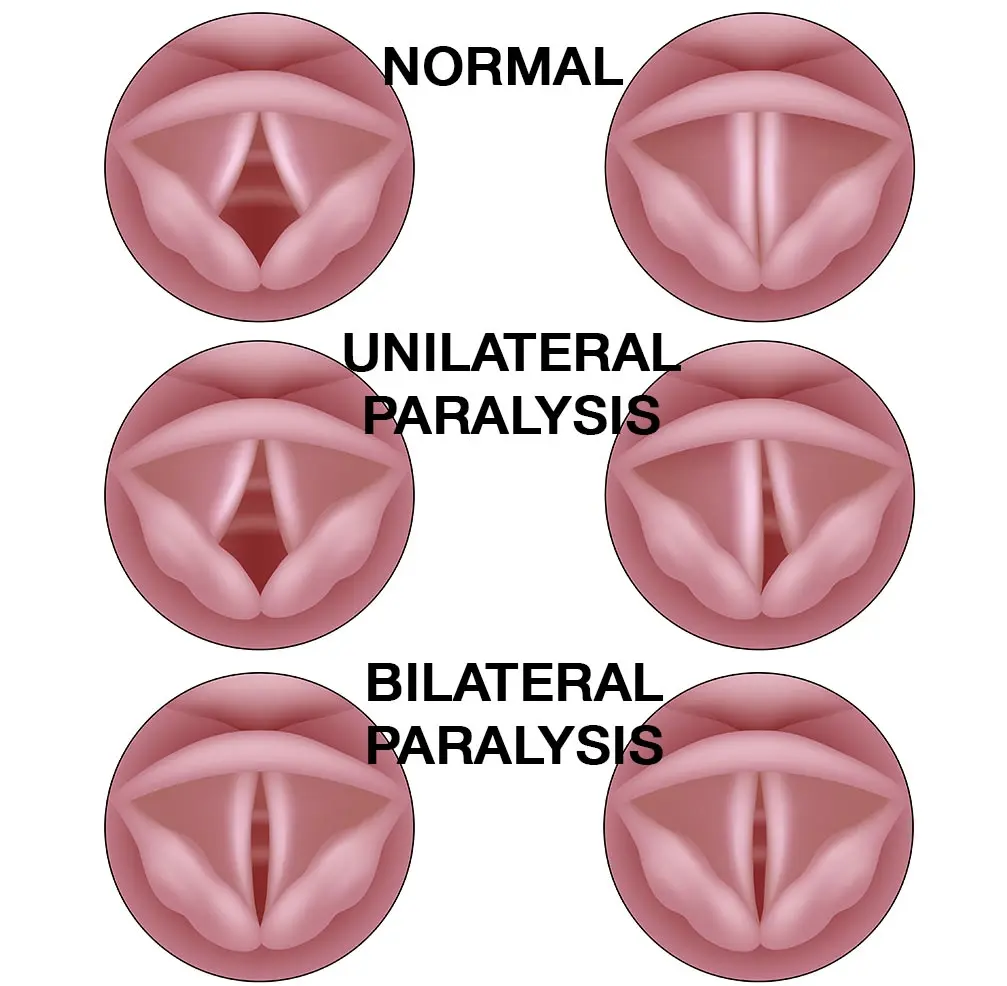
Laryngeal paralysis is when the arytenoid cartilages and vocal fold fail to abduct (open) during inspiration. Inspiration is then impaired and airflow resistance is increased, leading to arytenoid cartilage mucosal trauma, which further exacerbates the condition and establishes a vicious cycle.
It occurs when the nerves controlling the laryngeal muscles become paretic (weakened) or paralyzed (non-functional), preventing the muscles from properly pulling the airway open. When these muscles relax, the arytenoid cartilages and vocal fold collapse inward, blocking the airway. Rather than pulling open during inspiration, the walls of the airway are drawn into the passages and, in severe instances, pulled completely closed.
Laryngeal paralysis is a common cause of upper airway obstruction in large and giant breed dogs with 25% of all dogs being affected with some degree of laryngeal paralysis. Of all patients with upper airway obstruction, 5% of them are secondary to laryngeal paralysis.
Initially, it causes increased breathing noises. As it progresses, it can fully block the airway, leading to suffocation.
Laryngeal paralysis may affect either one side (unilateral) or both sides (bilateral), with bilateral presentation posing the greater severity.
Although this article focuses on acquired laryngeal paralysis, note that laryngeal paralysis stems from various causes, broadly classified into two categories: congenital (present from birth) and acquired (developing later in life).
Etiology
A lesion located anywhere from the nucleus ambiguus (medulla oblongata), cranial nerve X (vagus nerve), vagus nerve branches (recurrent laryngeal nerve, external branch of the cranial laryngeal nerve), or the cricoarytenoideus dorsalis muscle may result in acquired laryngeal paralysis in dogs.
Dogs affected by acquired laryngeal paralysis typically range from ~9.5 to 12.2 years of age, with a higher prevalence in males. Commonly affected breeds include the Labrador Retriever (which has an incidence of 35 to 73%), Golden Retriever, St. Bernard, Irish Setter, Afghan Hound, Chesapeake Bay Retriever, Great Dane, and Rottweiler.
Acquired laryngeal paralysis has been linked to a neuromuscular disease known as geriatric onset laryngeal paralysis and polyneuropathy (GOLPP); laryngeal paralysis frequently serves as an early indicator of GOLPP.
In addition to GOLPP, causes include neck or throat trauma leading to nerve dysfunction, space-occupying lesions or tumors, and endocrine diseases such as hypothyroidism and hyperadrenocorticism (Cushing’s disease). While these underlying endocrine diseases can be treated, therapy typically does not reverse or reduce the severity of the laryngeal paralysis.
The most common cause is idiopathic, a diagnosis of exclusion, making up 89% of cases, although neuromuscular disease is found in 22 to 56% of cases with 100% developing clinical signs by 1 year post-operatively.
Clinical Signs
With an insidious and progressive onset of months to years, the most common clinical signs include:
- Inspiratory laryngeal strider – 100% incidence
- Exercise intolerance, including excessive panting and trouble breathing – 90% incidence
- Change in the sound of the bark (dysphonia; hoarse or raspy bark) – 70% incidence
- Gagging and coughing (frequently associated with eating and/or drinking due to aspiration of food and/or fluid) – 60% incidence
- Expiratory laryngeal stridor – 55% incidence
In acute and severe cases, persistent excessive panting can induce laryngeal inflammation, resulting in respiratory distress. As a result, affected dogs may develop cyanosis (bluish mucous membranes) and may collapse.
Diagnostics
A diagnosis of laryngeal paralysis relies on evaluating a patient’s clinical signs alongside their medical history. Breathing difficulties and coughing frequently serve as the initial indicators of laryngeal paralysis.
Initial diagnostics may include blood work, urinalysis, thoracic radiographs, and ultrasonography, but in order to confirm the diagnosis of laryngeal paralysis, direct visualization of the arytenoid cartilages is needed.
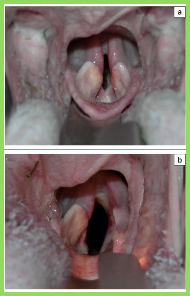
The most common method of diagnosis is direct visualization of the arytenoid cartilages via oral laryngoscopy (laryngoscopy per os). See Figure 1.
The degree of paralysis can be graded as follows:
- Grade 0 or 1: normal
- Grade 2: asynchronous full abduction occurs
- Grade 3: asynchronous incomplete abduction occurs
- Grade 4: marked asymmetry with no substantial movements or complete paralysis
Laryngoscopy is performed under sedation or anesthesia. The ideal protocol would maintain laryngeal function while simultaneously providing enough of a decrease in jaw tone to allow for effective and safe evaluation of laryngeal function by the surgeon and their equipment. Most protocols combine an anxiolytic, analgesic, and anesthetic.
Medical Treatment
Medical treatment targets the resolution or improvement of hypoxia, hyperthermia, excitement, and obesity.
For mild cases of laryngeal paralysis, this typically includes oxygen therapy, cooling measures, and medications such as sedatives and anti-inflammatories.
Emergency medical treatment, such as intubation and assisted ventilation, is indicated for the patient that already has or is about to experience cyanosis and collapse.
Note that for severe or congenital cases, the prognosis is guarded to poor without surgical intervention.
Surgical Treatment
Various surgical procedures are available depending on the severity of the patient’s laryngeal paralysis. Although surgery enhances airflow to the lungs and significantly boosts an affected dog’s quality of life, it cannot fully restore natural laryngeal function. Patients with laryngeal paralysis face a slightly higher risk of anesthetic complications. Additionally, because the larynx is held open after surgery, patients have an increased risk of aspiration pneumonia when eating or drinking.
The most commonly reported surgical treatment techniques include:
- Unilateral arytenoid cartilage lateralization (cricoarytenoid and/or thyroarytenoid laryngoplasty, also known as a laryngeal “tieback” procedure): this procedure helps maintain an open airway for easier breathing by using a suture to permanently pull open one side of the laryngeal wall, securing the collapsed cartilage to the side of the larynx
- Bilateral arytenoid cartilage lateralization
- Partial arytenoidectomy
- Partial laryngectomy
- Modified castellated laryngofissure
- Ventriculocordectomy
- Reinnervation of the laryngeal muscles
- Permanent tracheostomy tube placement
A combination of procedures or adjunct procedures (e.g. soft palate resection) may also be performed on a case-by-case basis.
The overall reported success rates resulting in resolution of clinical signs ranges from 65 to 100%, depending on case selection, surgical technique, surgeon experience, and anesthetic protocol.
There are several other procedures that are novel or experimental in nature. However, the unilateral arytenoid cartilage lateralization (or “tieback” procedure) is the most commonly used and probably the most effective procedure at this time. See figure 2 showing the intraoperative arytenoid cartilage position before correction (top picture) and after correction (bottom picture).
Bilateral arytenoid cartilage lateralization results in shorter survival times, higher risk of developing complications, and a higher risk of multiple complications due to the increased glottis diameter. Instead, the contralateral side is reserved for patients whose original unilateral arytenoid cartilage lateralization failed.
Suture failure can result from arytenoid cartilage fracture, arytenoid muscular process avulsion, suture breakage/rupture, and suture pull through. Factors that may lead to suture failure or cartilage fracture or avulsion include knot tightening, viscoelastic deformation of cartilage, patient age, and slowly progressive tissue tearing. Most failures are due to suture rupture.
Unilateral arytenoid cartilage lateralization can also be modified based on surgeon experience and the patient’s anatomy. These include any of the following individually or in combination:
- Cricothyroid joint versus cricoarytenoid joint disarticulations
- Cricoarytenoideus dorsalis muscle transection
- Interarytenoid sesamoid band transection
- Cricoarytenoid versus thyroarytenoid suture placement/lateralization
- Suture tension amount
Postoperative Care
Postoperatively, the patient is under continuous monitoring and held nil per os (NPO) for 12 to 24 hours while being supported with intravenous fluids. This is followed by offering 2 to 3 meatballs under supervision. Only soft canned food should be used without gravy or crumbs allowed. Ice cubes are then allowed for water intake. If tolerated, water is then offered. The patient should be closely observed for at least two weeks postoperatively.
Possible Surgical Complications
Complications associated with unilateral arytenoid cartilage lateralization include aspiration pneumonia, persistent coughing or a gagging after drinking (16 to 89%), revision surgery (10%), intramural hematoma formation, laryngeal webbing, seroma formation, voice change, suture failure (4 to 8%), gastric dilatation and volvulus (4%), respiratory distress (2 to 4%), acute postoperative respiratory distress, residual stridor (33%), arytenoid cartilage fragmentation, and death (3 to 19.3%). The complication rate ranges from 0 to 60%.
Factors that predispose a patient to complications include concurrent respiratory tract abnormalities, preoperative aspiration bronchopneumonia, concurrent esophageal disease, megaesophagus postoperatively, temporary tracheostomy tube placement, concurrent neoplastic disease, concurrent neurologic disease, body weight, age, surgeon experience, and surgeon familiarity with techniques.
The importance of concurrent neurologic disease on laryngeal paralysis development and its relationship to postoperative complications is becoming more obvious. The overall complication rate for dogs with neurologic comorbidities is 74%, compared with 32% for dogs without neurologic comorbidities. Those dogs with the presence of any neurologic comorbidity is associated with a 4-fold increase in complications and an 8-fold risk of recurring complications. The incidence of death in cases with both pneumonia and megaesophagus found postoperatively is 80%.
Aspiration pneumonia is the most common complication. Aspiration pneumonia is a lifelong concern in these patients as 0 to 33% of cases developed pneumonia postoperatively at some point with 0 to 14% resulting in death. The incidence of aspiration pneumonia after left-sided unilateral arytenoid lateralization in dogs at 7 days, 6 months, 1 year, 29 months, 3 years, and 4 years post-operatively is 4%, 5.1%, 2.5 to 18.6%, 4%, 31.8%, and 31.8%, respectively.
Pneumonia is suspected to occur secondary to aspiration as a result of either pharyngeal dysfunction (neuromuscular disease, dysphagia, and megaesophagus) or overcorrection of the rima glottidis opening beyond the edges of epiglottis. Preexisting aspiration pneumonia does not appear to be associated with an increased risk for development of aspiration pneumonia after surgery. In fact, clinical signs of pneumonia are not seen postoperatively if clinical signs for pneumonia were present preoperatively.
Surgical Survival Time and Rates
This surgery also carries the longest survival time ranging from 1 to 5 years with the overall median survival ranging from 12 to 60 months and the mean survival being approximately 15 months. By 4, 5, and 7 years postoperatively 70%, 70%, and 75% of cases are alive, respectively. The median survival rate is 8.5 months and the average survival rate is 11.8 months post-operatively in those dogs over 7 years of age. Death due to respiratory disease is seen in 14% of cases. Dogs that undergo surgical correction have a 2.6-fold reduction in the hazard of death.
Clinical signs resolve and good long-term respiratory function is seen in 82 to 100% of cases. Improvement in quality of life is seen in 90% of cases. Owner assessment of patient quality of life increases immediately postoperatively and until death, when compared with preoperative assessments. Owner satisfaction with large patients by six months postoperatively is 93%. Overall, 73 to 91% of owners are satisfied and 97% deem the surgery a success. The majority of patients appear to be able to resume normal activity.
Supportive Care
Supportive care includes preventing exposure to hot environments, avoiding strenuous exercise, maintaining ideal body weight, and using a harness instead of a collar.
References
- Andrade N, Kent M, Howerth EW, Radlinsky MG. Evaluation of Pharyngeal Function in Dogs with Laryngeal Paralysis Before and After Unilateral Arytenoid Lateralization. Vet Surg. 2015 Nov;44(8):1021-8.
- Bahr KL, Howe L, Jessen C, Goodrich Z. Outcome of 45 dogs with laryngeal paralysis treated by unilateral arytenoid lateralization or bilateral ventriculocordectomy. J Am Anim Hosp Assoc. 2014 Jul-Aug;50(4):264-72.
- Bookbinder LC, Flanders J, Bookbinder PF, Harvey HJ, Barry JS, Cheetham J. Idiopathic Canine Laryngeal Paralysis as One Sign of a Diffuse Polyneuropathy: An Observational Study of 90 Cases (2007-2013). Vet Surg. 2016 Feb;45(2):254-60.
- Cuddy LC, Case JB, Ellison GW, Covey JL. Video-assisted unilateral cricoarytenoid laryngoplasty in 14 dogs with bilateral idiopathic laryngeal paralysis. Vet Q. 2013 December;33(4):181-5.
- Greenberg MJ, Bureau S, Monnet E. Effects of suture tension during unilateral cricoarytenoid lateralization on canine laryngeal resistance in vitro. Vet Surg. 2007 Aug;36(6):526-32.
- Hardie RJ. Translaryngeal percutaneous arytenoid lateralization technique in a canine cadaveric study. J Vet Emerg Crit Care (San Antonio). 2016 Aug 27.
- Kitshoff A ,Van Goethem B, Stegen L, Vandekerckhov P, de Rooster H. Laryngeal paralysis in dogs: an update on recent knowledge. J S Afr Vet Assoc. 2013 Apr 5;84(1):E1-9.
- Milovancev M, et al. Vet Surg. Effect of Metoclopramide on the Incidence of Early Postoperative Aspiration Pneumonia in Dogs with Acquired Idiopathic Laryngeal Paralysis. 2016 Jul;45(5):577-81.
- Monnet E. Surgical Treatment of Laryngeal Paralysis. Vet Clin North Am Small Anim Pract. 2016 Jul;46(4):709-17.
- Nomura K, et al. Bilateral motion restored to the paralyzed canine larynx with implantable stimulator. Laryngoscope. 2010 December;120(12):2399-409.
- Paniello RC, Park AM, Bhatt NK, Al-Lozi M. Recurrent laryngeal nerve recovery patterns assessed by serial electromyography. Laryngoscope. 2016 Mar;126(3):651-6.
- Puram SV, et al. Vocal cord paralysis predicted by neural monitoring electrophysiologic changes with recurrent laryngeal nerve compressive neuropraxic injury in a canine model. Head Neck. 2016 Apr;38.
- Radlinsky MG1, Williams J, Frank PM, Cooper TC. Comparison of three clinical techniques for the diagnosis of laryngeal paralysis in dogs. Vet Surg. 2009 Jun;38(4):434-8.
- Shipov A, Israeli I, Weiser M, Kelmer E, Klainbart S, Milgram J. Minimally invasive unilateral arytenoid lateralization in dogs: A cadaveric study. Vet Sci. 2015 October;102:112-7.
- Stanley BJ, Hauptman JG, Fritz MC, Rosenstein DS, Kinns J. Esophageal dysfunction in dogs with idiopathic laryngeal paralysis: a controlled cohort study. Vet Surg. 2010 Feb;39(2):139-49.
- Stegen L, Kitshoff AM, Van Goethem B, Vandekerckhove P, de Rooster H. Intralaryngeal thyroarytaenoid lateralisation using the Fast-Fix 360 system: a canine cadaveric study. Vet Rec Open. 2015 Jul 27;2(1).
- Thieman KM, Krahwinkel DJ, Sims MH, Shelton GD. Histopathological confirmation of polyneuropathy in 11 dogs with laryngeal paralysis. J Am Anim Hosp Assoc. 2010 May-Jun;46(3):161-7.
18, von Pfeil DJ, Edwards MR, Déjardin LM. Less invasive unilateral arytenoid lateralization: a modified technique for treatment of idiopathic laryngeal paralysis in dogs: technique description and outcome. Vet Surg. 2014 Aug;43(6):704-11.
- Weinstein J, Weisman D. Intraoperative evaluation of the larynx following unilateral arytenoid lateralization for acquired idiopathic laryngeal paralysis in dogs. J Am Anim Hosp Assoc. 2010 Jul-Aug;46(4):241-8.
- Wen W, Sun G, Sun B, Liu C, Zhang M. Modified thyroplasty for unilateral vocal fold paralysis using an adjustable titanium implant. Eur Arch Otorhinolaryngol. 2015 Mar;272(3):517-22.
- Wilson D, Monnet E. Risk factors for the development of aspiration pneumonia after unilateral arytenoid lateralization in dogs with laryngeal paralysis: 232 cases (1987-2012). J Am Vet Med Assoc. 2016 Jan 15;248(2):188-94.
- Xu W, Han D, Hu H, Fan E. Characteristics of experimental recurrent laryngeal nerve surgical injury in dogs. Ann Otol Rhinol Laryngol. 2009 Aug;118(8):575-80.
- Zealear DL, et al. Stimulation of denervated muscle promotes selective reinnervation, prevents synkinesis, and restores function. Laryngoscope. 2014 May;124(5):E180-7.
- Zealear DL, et al. Rehabilitation of bilaterally paralyzed canine larynx with implantable stimulator. Laryngoscope. 2009 Sep;119(9):1737-44.
25. Martin-Flores M, Sakai DM, Campoy L, Cheetham J. A model of transient laryngeal hemiplegia in dogs through conduction blockade of the recurrent laryngeal nerve. Am J Vet Res. 2022 Jul 30;83(10):ajvr.22.04.0073. doi: 10.2460/ajvr.22.04.0073. PMID: 35895757; PMCID: PMC11753453.
26. Shi MX, Lan LL, Zhuang PY. Study on the Vocal Fold Movement Characteristics of Canine Models With Nerve Paralysis Caused by Different Site Injuries. World J Otorhinolaryngol Head Neck Surg. 2025 Mar 5;11(4):578-587. doi: 10.1002/wjo2.70005. PMID: 41477131; PMCID: PMC12753209.
27. Cai J, Kim YJ, Xu X, Ma Y, Scholp A, Jiang JJ, Liu T, Zhuang P. To Explore the Changes and Differences of Microstructure of Vocal Fold in Vocal Fold Paralysis and Cricoarytenoid Joint Dislocation by Diffusion Tensor Imaging. J Voice. 2023 Mar;37(2):187-193. doi: 10.1016/j.jvoice.2020.12.016. Epub 2020 Dec 31. PMID: 33388227.
28. Shelton GD, Carpentier MC, Kimura YM, Guo LT, Minor KM. A CNTNAP1 Missense Variant Associated With Laryngeal Paralysis and Polyneuropathy in Young Great Dane Dogs. J Vet Intern Med. 2025 Jul-Aug;39(4):e70185. doi: 10.1111/jvim.70185. PMID: 40622077; PMCID: PMC12231043.
29. Letko A, Minor KM, Friedenberg SG, Shelton GD, Salvador JP, Mandigers PJJ, Leegwater PAJ, Winkler PA, Petersen-Jones SM, Stanley BJ, Ekenstedt KJ, Johnson GS, Hansen L, Jagannathan V, Mickelson JR, Drögemüller C. A CNTNAP1 Missense Variant Is Associated with Canine Laryngeal Paralysis and Polyneuropathy. Genes (Basel). 2020 Nov 27;11(12):1426. doi: 10.3390/genes11121426. PMID: 33261176; PMCID: PMC7761076.
30. Srikanth K, von Pfeil DJF, Stanley BJ, Griffitts C, Huson HJ. Genome Wide Association Study with Imputed Whole Genome Sequence Data Identifies a 431 kb Risk Haplotype on CFA18 for Congenital Laryngeal Paralysis in Alaskan Sled Dogs. Genes (Basel). 2022 Oct 6;13(10):1808. doi: 10.3390/genes13101808. PMID: 36292693; PMCID: PMC9602090.
31.Jahns H, Vernau KM, Nolan CM, O’Neill EJ, Shiel RE, Shelton GD. Polyneuropathy in Young Siberian Huskies Caused by Degenerative and Inflammatory Diseases. Vet Pathol. 2020 Sep;57(5):666-674. doi: 10.1177/0300985820934112. Epub 2020 Jun 24. PMID: 32578500.
32. Asorey Blazquez C, Jolly Frahija I, Smith A, Miller R, Seth M, Garcia Manzanilla E, Valls Sanchez F. Retrospective Study of Chronic Coughing in Dogs in a Referral Centre in the UK: 329 Cases (2012-2021). Animals (Basel). 2025 Jan 17;15(2):254. doi: 10.3390/ani15020254. PMID: 39858253; PMCID: PMC11758286.
33. Anderson RS, Chu AK, Rylander H, Binversie EE, Duncan ID, Baker L, Salamat S, Patterson MM, Gruel J, Kohler NL, Kearney HK, Ale SM, Momen MM, Muir P, Svaren JP, Johnson R, Sample SJ. Pathologic classification of a late-onset peripheral neuropathy in a spontaneous Labrador retriever dog model. J Comp Neurol. 2024 Mar;532(3):e25596. doi: 10.1002/cne.25596. PMID: 38439568; PMCID: PMC10914337.
34. Madden M, Liatis T, Llanos C, Dancer S, Alvarez P, Tayler S, Hardas A, De Decker S. Jugular Foramen Syndrome: Concurrent Neurological Deficits, Advanced Imaging Findings, Underlying Diagnoses, and Outcomes in 14 Dogs (2016-2024). J Vet Intern Med. 2025 May-Jun;39(3):e70088. doi: 10.1111/jvim.70088. PMID: 40298565; PMCID: PMC12038936.
35. Touzet C, Hahn H, Gomes E, Bismuth C, Le Boedec K. Assessment of survey radiography as a method of diagnosing bilateral laryngeal paralysis in dogs. Vet Radiol Ultrasound. 2023 Mar;64(2):183-193. doi: 10.1111/vru.13192. Epub 2022 Dec 2. PMID: 36458646.
36. Natsume RE, Quina MT, Arble JB, Frederick SW, Drenning LE. Radiographic tracheal and carina distension is associated with diagnosis of laryngeal paralysis in dogs. J Am Vet Med Assoc. 2025 Jun 13;263(12):1540-1545. doi: 10.2460/javma.25.03.0173. PMID: 40523629.
37. Pan PC, Savidge C, Amsellem P, Hamilton S. Comparison between propofol and alfaxalone anesthesia for the evaluation of laryngeal function in healthy dogs utilizing computerized software. PLoS One. 2022 Jul 5;17(7):e0270812. doi: 10.1371/journal.pone.0270812. PMID: 35789223; PMCID: PMC9255722.
38. Perino JH, Patterson M, Momen M, Borisova M, Heslegrave A, Zetterberg H, Gruel J, Binversie EE, Anderson RS, Rylander H, Cameron S, Miranda B, Conidi G, Svaren J, Sample SJ. Neurofilament light chain concentration does not correlate with disease status in Labrador Retrievers affected with idiopathic laryngeal paralysis. Am J Vet Res. 2024 Feb 23;85(4):ajvr.23.12.0292. doi: 10.2460/ajvr.23.12.0292. PMID: 38382190; PMCID: PMC11059241.
39. Kapaldo N, McMurphy R, Hodgson D, Roush J, Berke K, Klocke E. Laryngeal function in normal dogs administered isoflurane following partial clearance of alfaxalone or propofol. Vet Anaesth Analg. 2021 Jul;48(4):493-500. doi: 10.1016/j.vaa.2021.03.009. Epub 2021 Apr 1. PMID: 33941487.
40. Ranninger E, Kantyka M, Bektas RN. The Influence of Anaesthetic Drugs on the Laryngeal Motion in Dogs: A Systematic Review. Animals (Basel). 2020 Mar 22;10(3):530. doi: 10.3390/ani10030530. PMID: 32235700; PMCID: PMC7143878.
41. Rishniw M, Sammarco J, Glass EN, Cerroni B. Effect of doxepin on quality of life in Labradors with laryngeal paralysis: A double-blinded, randomized, placebo-controlled trial. J Vet Intern Med. 2021 Jul;35(4):1943-1949. doi: 10.1111/jvim.16162. Epub 2021 May 17. PMID: 33998727; PMCID: PMC8295677.
42. Heaton JT, Kobler JB, Otten DM, Tynan MA, Petrillo RH, Ottensmeyer MP, Slate AR, Hillman RE, Zeitels SM. Electrical Stimulation of Vocal Fold Adduction Triggered by Laryngeal Electromyography Using a Custom Implant. J Speech Lang Hear Res. 2023 Dec 11;66(12):4812-4827. doi: 10.1044/2023_JSLHR-23-00377. Epub 2023 Nov 16. PMID: 37971489.
43. Paniello RC, Brookes S, Zhang H, Halum S. Enhanced Abductor Function in Bilateral Vocal Fold Paralysis with Muscle Stem Cells. Laryngoscope. 2024 Jan;134(1):324-328. doi: 10.1002/lary.30885. Epub 2023 Jul 18. PMID: 37462328.
44. Ricart MC, Rodríguez SM, Duré RM. Laryngeal stent for acute and chronic respiratory distress in seven dogs with laryngeal paralysis. Open Vet J. 2020 Apr;10(1):4-10. doi: 10.4314/ovj.v10i1.2. Epub 2020 Jan 16. PMID: 32426250; PMCID: PMC7193885.
45. Jeong SW. Silicone stenting as an emergency option for treatment of canine laryngeal paralysis. J Vet Sci. 2022 Jul;23(4):e69. doi: 10.4142/jvs.22199. PMID: 35920127; PMCID: PMC9346524.
46. Théron ML, Lahuerta-Smith T. Laryngeal silicone stent as a treatment option for laryngeal paralysis in dogs: a preliminary study of 6 cases. J Vet Sci. 2022 Jul;23(4):e58. doi: 10.4142/jvs.22068. PMID: 35920122; PMCID: PMC9346520.
47. David M, Giraud L, Dumont R, Gibert S, Dunié-Mérigot A, Boiron L, Sériot P, Jolivet F. Silicone stent placement for laryngeal diseases: a retrospective study of 14 dogs (2022-2025). J Small Anim Pract. 2026 May 24. doi: 10.1111/jsap.70143. Epub ahead of print. PMID: 42178139.
48. Li M, Chen M, Song W, Zheng H, Zhu W, Zhu M, Wang W, Song X. Synchronous Regeneration: Key Mechanism for Selective Reinnervation in Bilateral Vocal Fold Paralysis. Laryngoscope. 2026 Mar;136(3):1293-1301. doi: 10.1002/lary.70224. Epub 2025 Oct 29. PMID: 41159661; PMCID: PMC13001693.
49. Drudi D, Lisi MLP, Sommaruga P, Chiti LE, Massari F. Comparison of immediate and short-term outcomes of cricoarytenoid and thyroarytenoid lateralization in dogs with idiopathic laryngeal paralysis. Vet Surg. 2022 Apr;51(3):482-488. doi: 10.1111/vsu.13778. Epub 2022 Feb 2. PMID: 35107177.
50. MacGillivray KE, Bellefeuille SD, Hoffmann DE, St Germaine LL. Effects of a novel, 3D printed bilateral arytenoid abductor on canine laryngeal airway resistance ex vivo. BMC Vet Res. 2022 May 20;18(1):193. doi: 10.1186/s12917-022-03263-y. PMID: 35596179; PMCID: PMC9121604.
51. Shubert MP, Ganjei JB. Outcome following elective unilateral arytenoid lateralization performed in an outpatient manner is comparable to hospitalization for dogs with laryngeal paralysis. J Am Vet Med Assoc. 2023 May 23;261(9):1-6. doi: 10.2460/javma.23.02.0121. PMID: 37225155.
52. Sobczak GG, Morrison RA, Zhang L, Finnegan PR, Johnson B, Dong E, Egan B, Bhatt A, Voytik-Harbin SL, Halum S. Regenerative Injection Laryngoplasty With Scaffold-Forming Oligomer and Autologous Motor Endplate-Expressing Cells: A 6-Month Porcine Study. Otolaryngol Head Neck Surg. 2026 Aug 17. doi: 10.1002/ohn.70403. Epub ahead of print. PMID: 42606116.